
Clinical trials count on more than statistics
The rush to find treatments for COVID-19 led to a badly flawed clinical trial influencing medical treatment worldwide. What went…
General science


Spice up your physics lessons and show your students the tremendous impact of physics research on medical innovations.
In school, science is often taught in separate subjects. We have separate biology lessons, chemistry lessons, physics lessons, and so on. It’s natural to click with one subject and tune out on others. But these subtle preferences often decide which professional direction we pick later in our lives.
The world of natural sciences beyond school is not so neatly divided. At work, one might end up as a biologist on a team of computer scientists, or a physicist collaborating with biologists. It is very likely that we will need to understand concepts that are outside our area of expertise. The innovations in research and technology, as we experience them today, were only possible because we combined the forces of several disciplines. Interdisciplinarity is key!
Yet these connections are not always made explicit in classrooms. Showing students how ideas travel across subjects builds real-world understanding and motivation, especially for topics that may seem dry at first.
In the eye of the International Day of Medical Physics that is celebrated on the 7th of November, we decided to highlight the bridge between physics and medicine – a collaboration behind many clinical milestones!
We have curated a selection of Science in School articles that illustrate how physics, biology, and medicine work together to save lives. Use them as quick lesson starters, background reading, or discussion prompts. They give students concrete reasons why seemingly abstract topics matter. Attention in your lessons may rise once students see the connection to real life.
Target age: 14–19
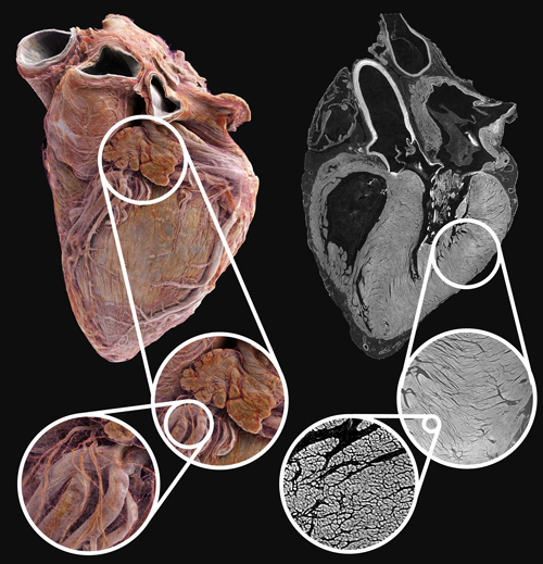
To obtain information about the heart tissue at the cellular level would usually require cutting into it, preparing thin slices, and examining them under the microscope. However, a new X-ray scanning technique is now able to generate a full 3D image of a human heart with details finer than a human hair and without any cutting required. This approach provides insight into the onset of numerous cardiovascular diseases and paves the way for new treatments. In the future, the method will be applied to other organs to create a comprehensive Human Organ Atlas. Whether you’re preparing an anatomy lesson or discussing X-rays, this article will capture your students’ attention.
Target age: 14–19
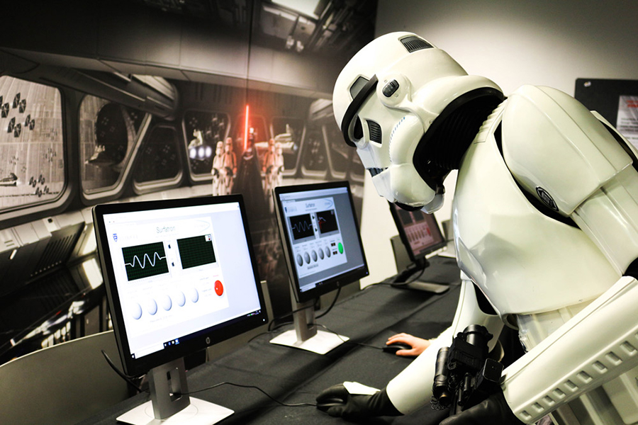
A proton torpedo, which you might know from the Star Wars movies, is science but definitely not fiction anymore. While it destroys the Death Star in the film, proton beams today offer a more precise and effective way to irradiate cancerous tissue in the human body compared with standard radiation therapies. The article draws a clear line from physics to medicine: from the discovery of the so-called Bragg peak of proton beams, through the invention of particle accelerators, to the translation of physics concepts and experiments into the clinic. It also shows the research still needed to further optimise proton-beam therapy. The article provides a perfect real-world example for your next lesson in particle physics.
Target age: 14–19
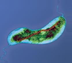
Besides protons, neutrons can also help to save lives, even though only in an indirect way. With small-angle neutron scattering (SANS), very small structures can be investigated. The article uses magnetotactic bacteria as an example: they contain chains of magnetic nanoparticles that function as an internal compass. These so-called magnetosomes are promising candidates for medical applications, for instance, to inspire the development of biomedical nanorobots that deliver drugs or perform minor procedures inside the body. To enable this, the structure and composition of magnetosomes must be fully understood, something SANS is well-suited to reveal. Talking about bacteria, imaging techniques, or neutrons, use this article to spotlight cutting-edge science.
Target age: 14–19
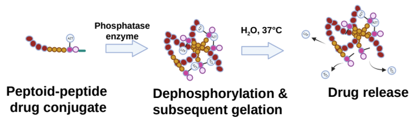
People infected with HIV need to take medicine every day for life to keep the virus under control. Everyone knows this can be easy to forget. Therefore, scientists are working on a delivery system that slowly and continuously releases HIV treatment into the bloodstream. It’s based on peptoids and peptides that form a hydrogel network around the medication after injection and then release the medication bit by bit into the bloodstream. But how is that studied? How do we know how peptoid–peptide hydrogels form and look like? How do researchers optimise it? It’s physics again! Scientists used SANS again to visualise these gel networks at the nanoscale. This article complements many school lessons on topics such as human metabolism, viruses, therapeutic windows, and neutrons. It also complements the aforementioned articles, which highlight SANS technology.
Target age: 14–19
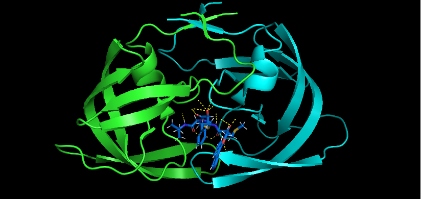
Another neutron-based technique used to study how a drug interacts with its target molecules is neutron crystallography. Many medicines work by increasing, decreasing, activating, or blocking the activity of specific biomolecules. To optimize these interactions, we need techniques that let us observe target molecules at an atomic scale. Neutron crystallography is especially suited for this since neutrons can determine the positions of all atoms, including hydrogens, something that normal X-ray crystallography often struggles to do. The article explains how this technique works and how it is helping to improve HIV therapy. Once again, a clear example that atomic-scale physics can drive life-saving medicine.
Target age: 14–19
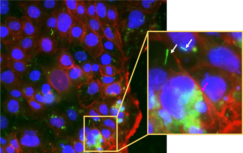
How do physical concepts such as radiation, absorption, and emission shape research on colorectal cancer? Colorectal cancer (cancer in the human gut) has been linked to an imbalanced microbiome in which harmful microbes overgrow. But whether this bacterial imbalance is a cause or a consequence of gut cancer remains unclear. To answer that, researchers use fluorescence microscopy. By tagging different bacterial groups with fluorescent probes, researchers can make them glow in distinct colours, revealing which bacteria are present, how they are distributed across healthy gut and tumour tissue, and whether they attach to or invade gut cells. Explore more about the physics behind fluorescence microscopy and how it untangles the microbiome-cancer puzzle in the article.
[1] Brunet J et al. (2024) Multidimensional Analysis of the Adult Human Heart in Health and Disease Using Hierarchical Phase-Contrast Tomography. Radiology 312: 1527–1315. doi: 10.1148/radiol.232731
[2] Coutler SM et al. (2024) In Situ Forming, Enzyme-Responsive Peptoid-Peptide Hydrogels: An Advanced Long-Acting Injectable Drug Delivery System. Journal of the American Chemical Society 146: 21401–21416. doi: 10.1021/jacs.4c03751
[3] Adachi M et al. (2009) Structure of HIV-1 protease in complex with potent inhibitor KNI-272 determined by high-resolution X-ray and neutron crystallography. Proceedings of the National Academy of Sciences 106 (12): 4641-4646. doi: 10.1073/pnas.0809400106
The rush to find treatments for COVID-19 led to a badly flawed clinical trial influencing medical treatment worldwide. What went…
Sarah Garner and Rachel Thomas consider why well-designed and properly analysed experiments are so important when testing how effective a medical…


When your doctor prescribes you a tablet and you get better, was it really the drug or could it have been the colour of the tablet? Andrew Brown…


