On the trail of viruses: understanding virus research Teach article

DNA-based methods such as PCR, gel electrophoresis and bioinformatics, provide a practical insight into virus research for students – applicable to COVID-19, bird flu and others.

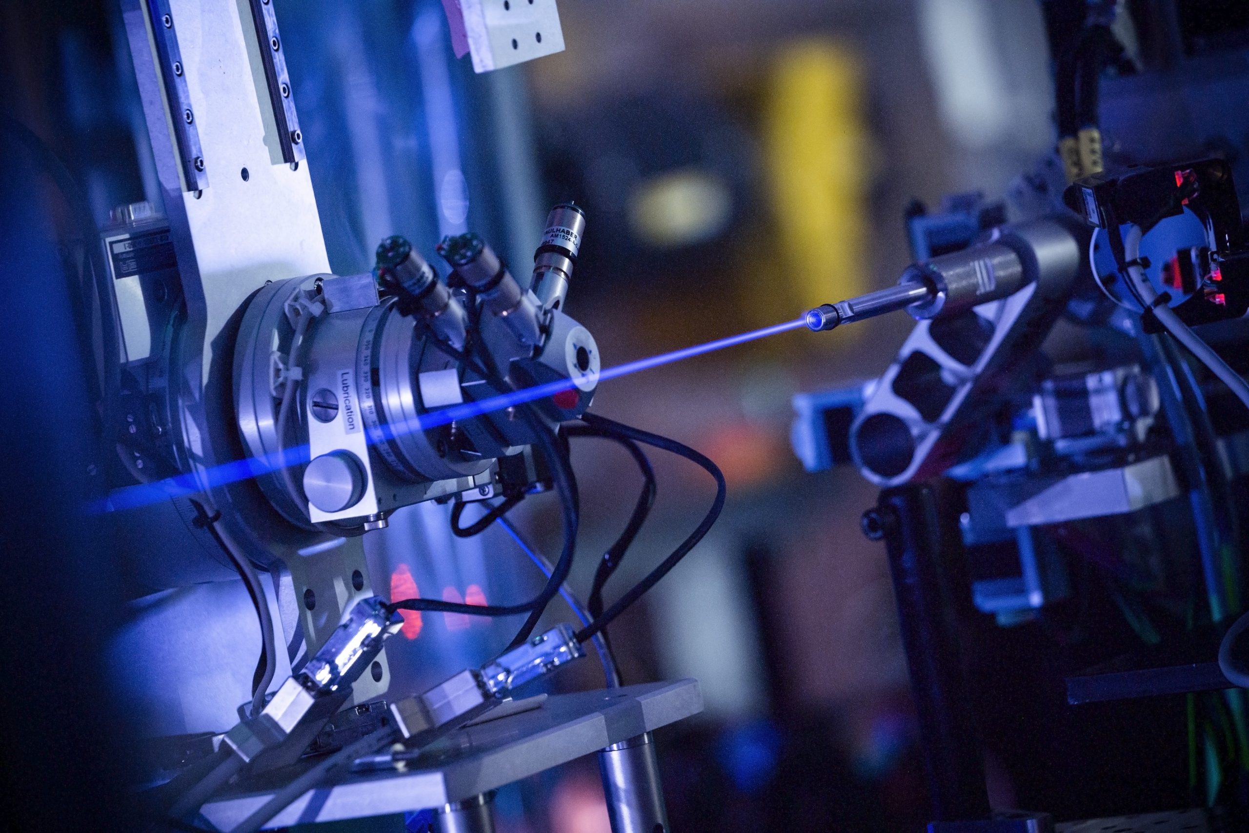
Viral pathogens like SARS-CoV-2, bird flu and Ebola pose recurring global challenges requiring rapid scientific response.[1] In this unit, students apply molecular biology methods to compare virus variants at the DNA level using a bioinformatics software. They explore essential questions: Why are primers needed? What makes mutations dangerous?[2] How does bioinformatics aid vaccine development?[3,4] Activities connect to real research at the European XFEL (X-Ray Free-Electron Laser Facility), where scientists use ultra-short X-ray pulses to visualise SARS-CoV-2 proteins.[5] This demonstrates the modern science of being networked, data-driven and socially relevant. As well as acquiring specialised knowledge, students develop scientific thinking skills such as precise observation, logical reasoning, and critical evaluation using methods applicable across viral diseases.[6]

Image: DVisions/Adobe Stocks, Standard License

Activity 1: Short movie about virus research

A short, visually impressive video gives the students an initial insight into modern virus research. The focus is on how large-scale scientific research facilities, such as the European XFEL, help to visualise new virus structures, which form the basis for vaccine development, drug research and pandemic preparation.

Materials

Procedure

  1. Start with a short thematic introduction (2–3 min), asking the following questions:
    a) How do you investigate something that you can barely see with a microscope?
    b) What role does physical technology play in biology?
  2. Watch the video together (approx. 10 min) (Note: The video can also be watched individually in advance or at home).
  3. Work on Worksheet A in individual or partner work.

Results/discussion

The solutions for each task can be found in the supplemental materials Solution worksheet A.

As a teacher, you will moderate a joint evaluation in a plenary session:

  • What surprises you about the connection between physics and biology?
  • Why does modern biology need international research institutions?
  • Could such methods also help with other viruses?

A detailed explanation for each of these questions can be found in the Discussion answers.

Learning objectives: 

  • Students understand that virus research is highly interdisciplinary.
  • They recognise that structural elucidation is of central importance for diagnostics and therapy.
  • They get to know an example of modern research infrastructure.

Activity 2: DNA sequence analysis – Understanding mutations in SARS-CoV-2

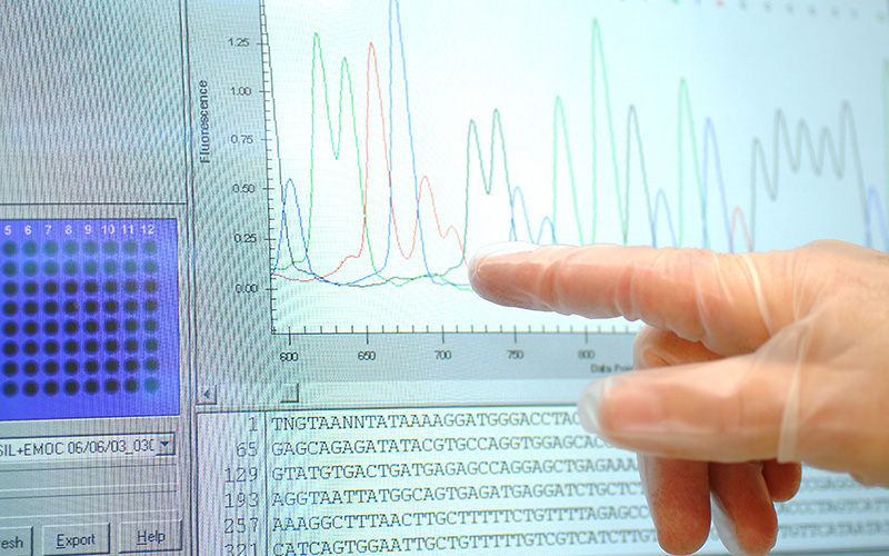
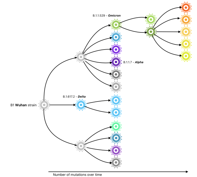
This lesson introduces students to DNA sequence analysis using real SARS-CoV-2 variants. Using the free MEGA software, students will learn to identify point mutations by comparing spike protein gene sequences from the wild type, Delta and Omicron variants. This hands-on activity demonstrates how mutations arise, how they’re detected and their potential effects on the protein structure and function. Students will gain practical experience of how to use bioinformatics tools, while developing an understanding of the evolutionary processes that drive viral adaptation.

DNA structure and base pairing
Image courtesy of the author

Learning Objectives:

  • Identify three types of point mutations (substitution, deletion, insertion)
  • Use sequence alignment software to compare DNA sequences
  • Translate nucleotide sequences to amino acid sequences
  • Understand how mutations affect protein structure and viral evolution

Duration: 90–120 min

Material

Per student or pair:

Teacher preparation:

  • Install MEGA 11 on all computers 1–2 days before lesson.
  • Test the software and familiarise yourself with the interface.
  • Prepare the sequence.fas file with the wild type, Delta and Omicron sequences.
  • Print the worksheets and reference materials.
  • Review the mutation nomenclature (e.g., G102Y format).

Procedure

Phase 1: Software Introduction (15 min)

  1. Demonstrate how to open MEGA and how to load the sequence file (File → Open A File/Session).
  2. When prompted “Analyse or Align File?” select “Align”.
  3. Show students the initial unaligned sequences.
  4. Have students complete task 1: observing the start codon (ATG) and initial sequence structure.

Phase 2: Sequence alignment (20 min)

  1. Guide students through the alignment procedure: Alignment menu → “Align by MUSCLE (Codons)”.
  2. Accept the default parameters and select “Yes” when asked to remove gaps.
  3. Choose the “w/o gaps” option in the alignment window.
  4. Students complete task 2: comparing aligned vs unaligned sequences and identifying mutation examples.

Phase 3: Mutation investigation (35 min)

  1. Demonstrate navigation using the “Site #” field at bottom left.
  2. Explain codon positioning: divide the site number by 3 to determine the position within a codon.
  3. Students examine four specific mutation sites (205–210, 642–643, 1501–1503, 2041–2043).
  4. For each site, students identify:
    • which variant is mutated;
    • the type of point mutation;
    • the nucleotide changes;
    • the resulting amino acid changes (using codon table in Worksheet B).
  5. Verify results using the “Translated Protein Sequences” function.

Phase 4: Analysis and discussion (25 min)

  1. Students complete task 4, analysing:
    • deletion mutation Δ69–70 and PCR detection implications;
    • side chain properties in N501Y mutation;
    • effects of P681R (Delta) and P681H (Omicron) near cleavage sites.
  2. Discuss with the whole class the functional consequences of mutations.
  3. Explain the connection of mutations to selection pressure and viral evolution concepts.

Phase 5: Extension activities (homework)

  • Students may research artificial virus modifications (gain-of-function research).
  • Students may investigate the PCR testing methodology for COVID-19 detection.
Family tree of SARS-CoV-2 virus strains. The first known strain (B1 from Wuhan) is shown on the far left. It represents the starting point. The further you move to the right, the more mutation there are. Same colours stand for the same mutation. The strains Delta, Alpha and Omikron are highlighted in the family tree as they have appeared most frequently during the course of the pandemic
Image courtesy of the author

Results/Discussion

Students should identify silent, missense and nonsense mutations and recognise that missense mutations predominate in viable viral variants. The Δ69–70 deletion demonstrates how mutations affect diagnostic testing. The analysis of amino acid side chains (polar, nonpolar, charged) reveals how mutations alter protein interactions and potentially affect viral infectivity. The solutions for each task can be found in the supplemental materials in the Solution worksheet B.

Discussion points could be:

  • Why do certain mutations appear in multiple variants?
  • How does selection pressure drive mutation accumulation?
  • What role do amino acid properties play in protein function?
  • Why might some mutations increase viral transmission?

A detailed explanation for each of these questions can be found in the Discussion answers.

Evaluate the worksheets of your students for accuracy in mutation identification, codon translation, and understanding of structure-function relationships in proteins.

Activity 3: Primer design – a work sheet orientated task

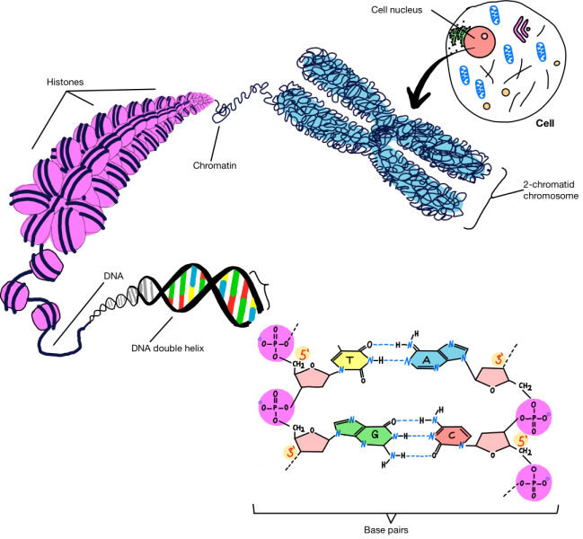
This exercise teaches students how to design DNA primers for PCR applications. Students will learn about DNA structure and base pairing rules as well as calculate primer specificity and melting temperatures using mathematical formulas.

Materials

  • Discussion answers
  • Calculator or computer
  • Pencil and paper
  • Optional: internet access to demonstrate the primer design with the software 

Procedure

Part 1: Understanding primer specificity

  1. Review with your students the DNA structure: four bases (A, T, G, C) with complementary pairing (A-T, G-C),
  2. Explain that primers are 15–30 nucleotides long and must bind specifically to the target DNA.
  3. Calculate possible combinations for different primer lengths using the formula 4n (where n is the number of nucleotides).
  4. Students complete task 1: Calculate combinations for 8-nucleotide and 17-nucleotide primers.
  5. Divide 3 billion (human genome base pairs) by your calculated combinations to find potential binding sites.
  6. Discuss why longer primers (17–28 nucleotides) are more specific.

Part 2: Calculating melting temperature

  1. Introduce the melting temperature (Tm) formula Tm = 4 × (G+C) + 2 × (A+T) [°C].
  2. Explain that annealing temperature (Ta) is 1–2°C below Tm.
  3. Students complete task 2: Calculate Tm for three given primer sequences.
  4. Count the G/C and A/T bases in each sequence.
  5. Apply the formula and determine the optimal annealing temperatures.
  6. Discuss how the GC content affects primer stability and why proper temperature selection prevents non-specific binding.

Results/Discussion

Students should find that 8-nucleotide primers have too many potential binding sites, while 17+ nucleotide primers are sufficiently specific. The solutions for each task can be found in the supplemental materials in the Solution worksheet C and the answers to the discussion points in the Discussion answer.

Acknowledgements

This article could not have been written without the funding of the Joachim Herz Foundation (https://www.joachim-herz-stiftung.de/en/), who funded the work of Anusha Veena and the materials used. I would like to especially thank Anusha Veena for her work and input in creating these activities together with Sarah Aretz and Arwen Cross from the European XFEL GmbH. 


References

[1] Ilyichev AA et al. (2020) mRNA technology as one of the promising platforms for the SARS-CoV-2 vaccine development. Vavilovskii Zhurnal Genet Selektsii 24: 802–807. doi: 10.18699/VJ20.676

[2] Mentes A et al. (2022) Identification of mutations in SARS-CoV-2 PCR primer regions. Sci Rep. 12:18651. doi: 10.1038/s41598-022-21953-3

[3] Franziska Hufsky et al. (2022) Computational strategies to combat COVID-19: useful tools to accelerate SARS-CoV-2 and coronavirus researchBriefings in Bioinformatics 22: 642–663. doi: 10.1093/bib/bbaa232

[4] García-Machorro J et al. (2022) The Advantage of Using Immunoinformatic Tools on Vaccine Design and Development for Coronavirus. Vaccines 10: 1844. doi: 10.3390/vaccines10111844

[5] Sebastian Günther et al.(2021) X-ray screening identifies active site and allosteric inhibitors of SARS-CoV-2 main protease. Science 372: 642-646. doi: 10.1126/science.abf7945

[6] Lewitter F, Bourne PE (2011) Teaching bioinformatics at the secondary school level. PLoS Comput Biol. 10: e1002242. doi: 10.1371/journal.pcbi.1002242

Resources

Institutions

Author(s)

Sebastian Frerichmann combines a strong foundation in molecular plant biology, shaped by phytopathology research at the University of Hamburg and a doctoral thesis at the Plant Breeding Institute Kiel. He is currently seconded to the Xcool Lab at European XFEL as a teacher, where he coordinates the molecular biology laboratory and advances interdisciplinary approaches in plant science and experimental methods.

License

CC-BY
Text released under the Creative Commons CC-BY license. Images and supporting materials: please see individual descriptions.

Related articles

Understand

Vaccines in the spotlight

Hit me with your best shot: Vaccines have taken centre stage in the COVID-19 pandemic. What are the different types and how do they…