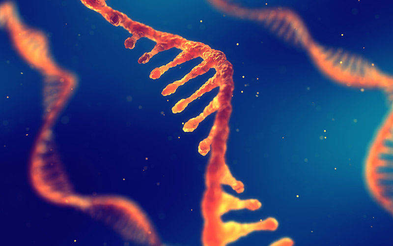
Not just a blueprint for proteins: the importance of non-coding RNAs
We all know that DNA → RNA → protein. But did you know that some genes don't encode proteins but rather RNAs with important cellular functions?

Take a virtual tour of The World of Molecular Biology to access awe-inspiring microscopy images and explore cutting-edge life science themes.
The World of Molecular Biology is a new exhibition at the European Molecular Biology (EMBL) headquarters in Heidelberg, Germany. It uses a combination of animation, images, games, and virtual reality to take visitors on a journey through the inner workings of life. A visit to the exhibition, with its many interactive elements, is a great opportunity to see the fundamental concepts of molecular biology, from genomes to ecosystems, in a different light.
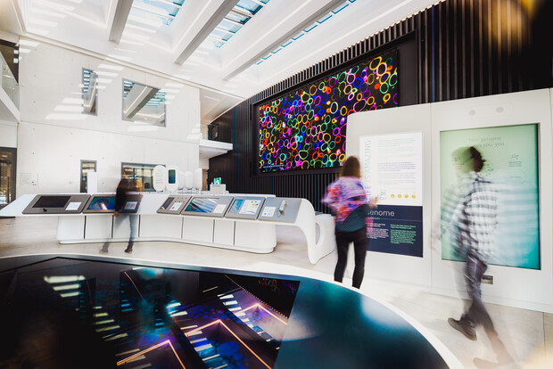
But schools don’t have to travel to where the exhibition is physically located to discover its awe-inspiring images and behind-the-scenes insights into lab life. Thanks to a novel virtual tour option, the World of Molecular Biology is accessible to anyone with a digital device. Taking this virtual tour can bring the following subjects to life:
The virtual tour works best as a visual exploration of the themes, and to access a range of informative videos with a huge amount of useful educational information. It doesn’t feature interactive content, but there are some virtual tour exclusives that are only available to digital visitors!
The virtual tour is accessible via a web browser. The Download button at the top of the page enables offline use.
Virtual visitors start the tour standing in front of the Imaging Centre, the newest building on EMBL’s Heidelberg campus. Clicking on ‘+’ reveals more information about a specific exhibit or element – in this case, a description of the building, including a video and website link.
Once inside, visitors can wander around the exhibition using the symbols on the floor (green circles and arrows navigate to the next point of interest), or by clicking on the left-hand panel to get to a specific section, marked with green, purple, and blue tabs. This panel also features a map of the exhibition, pinpointing the visitor’s location. The panel can be hidden and re-accessed by clicking on the black arrow.
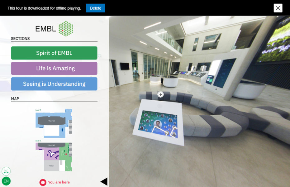
Clicking on the white ‘+’ symbol at each exhibit enables access to the related content, in the form of videos or slideshows (videos are indicated with a green circle around the +). Slideshows offer a glimpse of what would normally be an interactive exhibit (for example, a game); the slides can be navigated by clicking on the left and right arrows, or you can skip to a specific one using the grey circles below the image.
The World of Molecular Biology is divided into three sections: Spirit of EMBL (green tab) offers an introduction to EMBL’s people, history, mission, and values. The most extensive section, Life is Amazing (purple tab) takes visitors on a journey of scale, from the smallest elements of life (genes) to ecosystems. The third section, Seeing is Understanding (blue tab) explains light and electron microscopy techniques and highlights some of the special microscopes developed by and hosted in the EMBL Imaging Centre. It also gives an insight into how artificial intelligence (AI) aids in advancing life-sciences research.
The first exhibit features video interviews with nine EMBL employees in a variety of roles across six sites, ranging from EMBL’s Director General, Edith Heard, to research group leaders and a PhD student. These videos showcase different EMBL research fields, but also offer insights into diverse science careers that don’t fit the classic ‘scientist’ image, such as marine animal technician or mechatronics engineer, offering inspiration for the next generation.
In this section, the focus expands from the cellular level to planetary biology, with themes including molecular machines, cells, organisms, and ecosystems. Visitors can discover how much of their DNA is shared with a banana (more than they might expect!) and learn about how genes drive the processes of life. The Human Ecosystem section reveals how physical, biological, and social environments influence human health, and how vast databases can help scientists drill down into the detail of this complex web of influences. Visitors can also view content focused on genome packaging, flu virus replication, and the gut microbiome. Virtual-reality goggles link to footage showcasing ground-breaking new insights into cell division in fertilized mammalian embryos. This video highlights EMBL scientist Judith Reichmann’s 2018 discovery that maternal and paternal genomes are organized in the developing embryo via a dual-spindle mechanism.[1] The huge LED screen on the back wall displays amazingly detailed microscopy images. Clicking on this ‘Window of Wonder’ allows access to an eight-minute video loop.
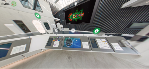
This section highlights the latest microscopy technologies, which allow scientists to observe things that have never been seen before. Cryo-electron microscopes use electron waves rather than light to enable atomic-level image acquisition. While working at EMBL, biophysicist Jacques Dubochet developed a technique to catch biological processes in action by rapidly freezing samples without damaging them, for which he and two other scientists received the 2017 Nobel Prize in Chemistry.[2] This discovery was central to the development of the cryo-electron microscope. Understanding protein structures in such detail gives us vital clues as to how molecules interact with each other to promote health or cause disease.
To maximize the clarity and resolution obtained using the cryo-electron microscopes, EMBL’s Imaging Centre is constructed around an insulated inner cryo-hall with its own separate foundations to protect the equipment from vibrations and ensure maximum sensitivity. The area is normally off-limits to visitors, but digital guests can access a virtual tour exclusive: footage of a scientist loading a sample into the cryo-electron microscope and starting the imaging process. To view this, follow the green arrows to the panel ‘Electron microscope for frozen samples’ and click on the window.
The Seeing with Light section features another exclusive virtual tour video, showcasing a light-sheet microscope. A technology first developed at EMBL, light-sheet microscopy enables live samples to be clearly imaged using low-intensity light ‘sheets’, causing minimal damage to cells. Hundreds of image slices are then collated to create 3D images and videos of cell-to-cell interactions. This is vividly highlighted in a video showing the development of a fruit fly from embryo to larva in real time.
Finally, around the corner, visitors reach two of the exhibition’s highlights: 3D imaging and AI. Two sets of virtual-reality goggles host videos on building 3D images and mapping the orchestra of proteins in a dividing cell. Directly opposite, the final exhibit introduces AI and its role in research and features interviews with Anna Kreshuk and Virginie Uhlmann, who are pushing the boundaries of life sciences with machine learning.
Visitors to the virtual tour often feel inspired to make the journey in real life. EMBL hosts free guided tours and linked workshops for schools, and has welcomed young learners from as far away as Greece, the Netherlands, and France. Teachers are invited to get in touch with the Science Education and Public Engagement (SEPE) office at EMBL to arrange a real-life tour, or book directly online.
EMBL is Europe’s leading laboratory for the life sciences. An intergovernmental organization supported by over 25 member states and operating across six sites in Europe, EMBL performs fundamental research in molecular biology, studying the story of life. Its research drives the development of new technology and methods in the life sciences, with the aim of sharing this knowledge for the benefit of society. The EMBL head office (along with The World of Molecular Biology exhibition) is in Heidelberg, Germany.
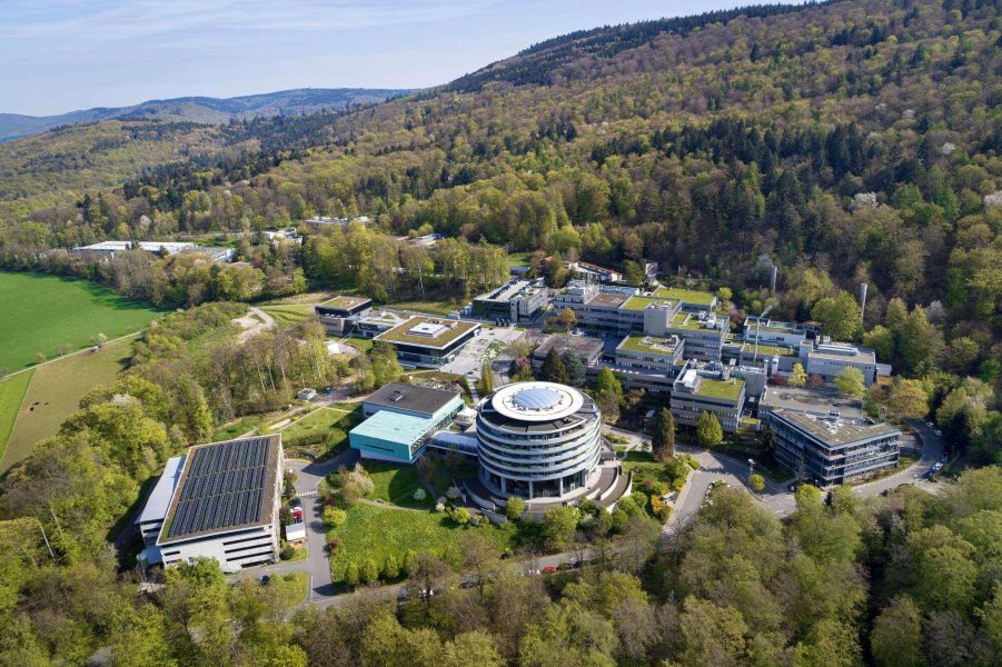
The World of Molecular Biology exhibition was developed and curated by EMBL’s Science Education and Public Engagement (SEPE) office, which leads the institute’s science education activities and coordinates its outreach programmes. These include public engagement events and training, free educational resources on subjects from Genomics to Ecology, teacher training for secondary school teachers keen to bring real science into the classroom, the Microscope in Action programme (where schools can lease fluorescence microscopes for free), the annual Insight lecture for young learners, and Ambassador programmes for teachers.
[1] Reichmann J et al. (2018) Dual-spindle formation in zygotes keeps parental genomes apart in early mammalian embryos. Science 361:189-193. doi: 10.1126/science.aar7462
[2] Jacques Dubochet, Joachim Frank, and Richard Henderson are awarded the Nobel Prize in Chemistry, 2017: https://www.nobelprize.org/uploads/2018/06/popular-chemistryprize2017.pdf

We all know that DNA → RNA → protein. But did you know that some genes don't encode proteins but rather RNAs with important cellular functions?
Why was a Nobel prize awarded for 'click chemistry'? Learn about the ground-breaking advance behind this simple-sounding…
The PDB Art project aims to make science more accessible and inspire young people to explore the beauty of proteins by bringing together art and…