The resting potential: introducing foundations of the nervous system Teach article

Simulate a neuron in the classroom.

issue38_membrane
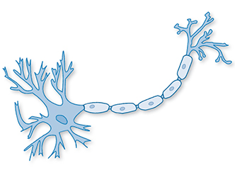
Image courtesy of Dhp1080;
image source: Wikimedia
Commons

The nervous system is not only fascinating but also probably one of the most complex topics in school biology lessons, particularly because working with real neurons is not feasible at school. In this article, we describe an activity that uses a cellophane membrane to explore how the resting potential is generated in a neuron. Suitable for students aged 16–19, the activity takes approximately 90 minutes.

An artificial membrane potential

To transfer information, neurons need to be able to generate and maintain a membrane potential: a voltage difference between the intracellular and extracellular media that is focused along the cell membrane. The voltage difference in an unexcited neuron is referred to as the resting potential. Stimulating this neuron can alter the resting potential, causing an action potential: the electrical impulse by which the neuron transmits information. Before the neuron can fire again, the resting potential needs to be re-established (figure 1). But how is the resting potential generated and maintained? The answer lies partly with the semi-permeable nature of the cell membrane.
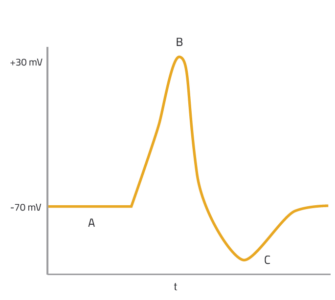
Figure 1: Voltage difference across the cell membrane over time, when a neuron is stimulated. A: the resting potential; B: an action potential; C: the resting potential is re-established; t: time.
Image courtesy of Nicola Graf

Among other constituents of the intracellular and extracellular media are dissolved ions including sodium (Na+), chloride (Cl), organic anions (A) and, most importantly, potassium (K+). Once a neuron has fired and the resting potential begins to be re-established, the concentration of K+ ions is higher inside the neuron than outside. Unlike most other ions, K+ ions can pass freely into and out of the cell, via specialised ion channels in the membrane. Driven by the concentration gradient, K+ ions diffuse out of the neuron, causing a net movement of positive charge (figure 2). This causes a voltage difference across the membrane, with the intracellular medium being more negatively charged than the extracellular medium. This is the resting potential, with a value of around –70 mV.

Figure 2: When the solutions on either side of a semi-permeable membrane have different concentrations (top), the distribution of negative and positive charges across the membrane becomes unbalanced (bottom), causing a voltage difference. Note that the concentration of K cations remains higher on one side of the membrane, due to their attraction to the anions, which are trapped by the membrane.
Image courtesy of Alexander Maar

Although there are additional factors involved in establishing the resting potential in a neuron, the combined contribution of the concentration gradient and the electrical properties of anions and cations can easily be demonstrated in the classroom using cellophane as a semi-permeable membrane, as described below.

Before the activity, it is useful to cover the basic principles of diffusion and cell membranes with your students. Instructions for hands-on activities involving the properties of the cell membrane and diffusion through membranes can be downloaded from the additional material sectionw1.

Materials

For each group of 2–4 students, you will need:

  • 300 ml 0.01 M potassium chloride (KCl) solution
  • 100 ml 0.1 M potassium chloride (KCl) solution
  • Distilled water
  • Voltmeter
  • Electrodes (chlorinated silver wire)
  • Glass bowl (200–300 ml)
  • Funnel
  • Cellophane wrapping
  • Rubber band
  • Clamp stand and three clamps
  • Two cables with crocodile clips
  • Pipettes
  • Scissors

Procedure

Before beginning the activity, discuss with your students how they think a voltage difference can occur in the cell, and which cell components are important in establishing it. Briefly introduce the resting potential. Then, ask your students to:
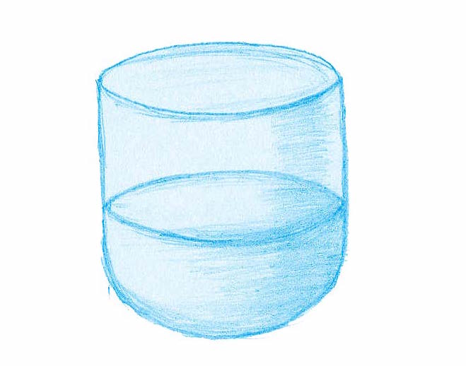
  1. Fill the glass bowl with about 200 ml of the 0.01 M potassium chloride (KCl) solution, which represents the extracellular medium of the membrane.
Image courtesy of Alexander Maar
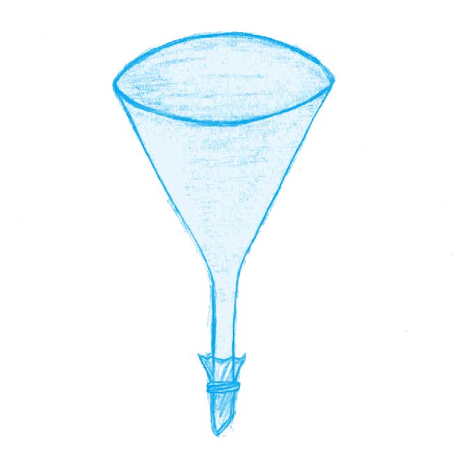
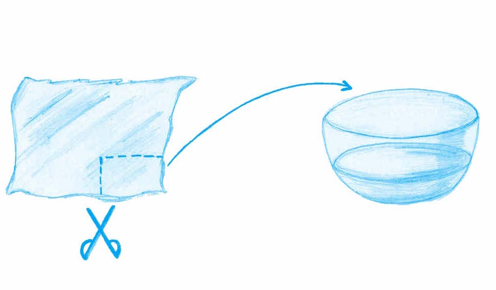
  1. Cut a piece of cellophane wrapping large enough to cover the base of the funnel, then rinse the cellophane in distilled water to make it more flexible. The cellophane serves as the semi-permeable membrane.
Image courtesy of Alexander Maar
  1. Wrap the piece of cellophane tightly around the base of the funnel and seal it with the rubber band.
Image courtesy of Alexander Maar
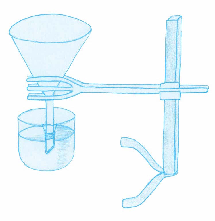
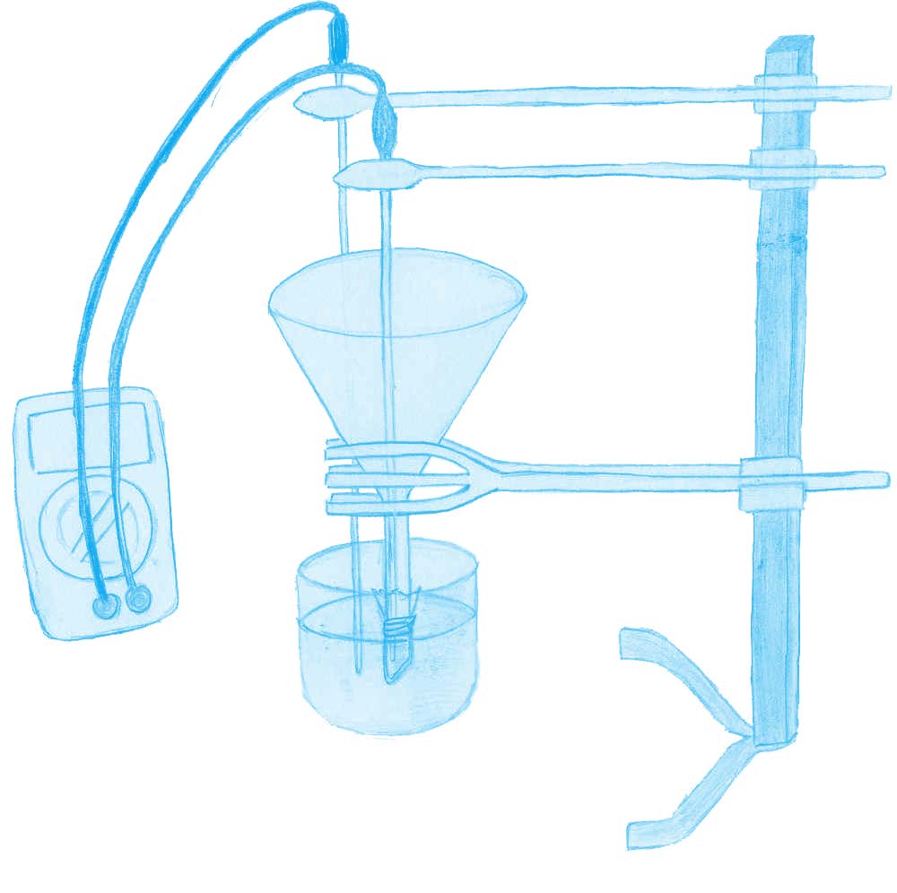
  1. Clamp the funnel to the clamp stand, submerging the base of the funnel in the KCl solution in the glass bowl.
Image courtesy of Alexander Maar
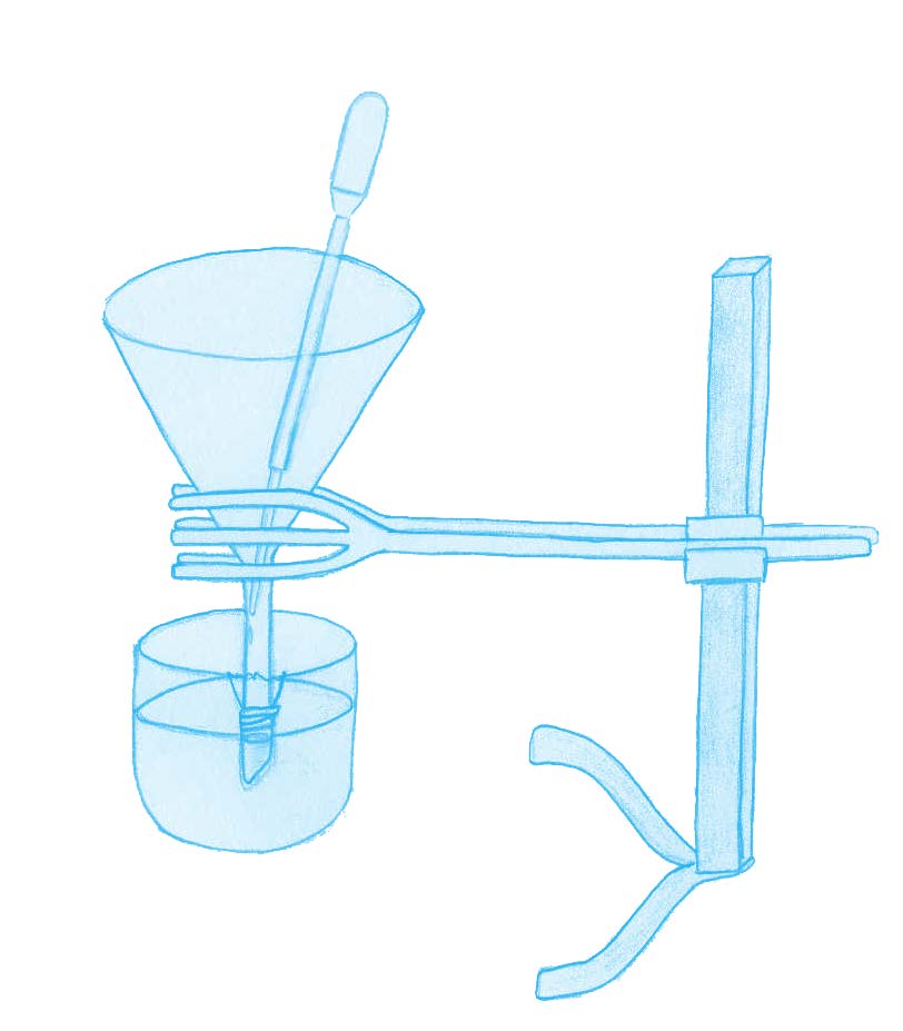
  1. Using a pipette, add 0.1 M KCl solution to the funnel until the liquid levels inside and outside the funnel are level. The solution inside the funnel represents the intracellular medium.
Image courtesy of Alexander Maar
  1. Attach the two electrodes to the voltmeter with crocodile clips. Using a clamp, place the electrode that is connected to the cathode of the voltmeter in the solution in the glass bowl. Supported by a further clamp, place the second electrode, connected to the anode, in the solution in the funnel.
Image courtesy of Alexander Maar

Discussion and further investigations

Ask your students:

  • What voltage do they predict that the voltmeter will show? Some students may think that it will be positive, as is an action potential. Ask them to set the voltmeter to approximately 200 mV.
  • Within about 10 seconds, the voltage will decrease, stabilising after about 5 minutes at about –50 mV to –60 mV.
  • What causes the voltage difference between two solutions? Why is the value negative? What would have happened if the solution in the glass bowl had been the more concentrated of the two?
  • Why do they think the membrane and the two solutions generated an unequal distribution of ions?

As in the real-life neuron, this experiment relies on two components: a concentration gradient and the semi-permeable properties of the cellophane wrapping. Like the membrane of a neuron, the cellophane is permeable to K+ ions but almost non-permeable to Cl ions. As a result, as in the neuron, there is a gradual, net diffusion of K+ ions out of the funnel (0.1 M KCl) and into the glass bowl (0.01 M KCl). If the electrodes are placed carefully, without puncturing the cellophane, the voltage of the solution in the funnel can be seen to become more negative. The initial setting of 200 mV on the voltmeter is arbitrary, to ensure that the final reading is similar to that of the real-life resting potential.

While realistic, this experiment is not a complete model of how the resting potential is established and maintained. In a neuron, the extracellular and intercellular media contain more than just K+ and Cl ions, and there are additional mechanisms that determine the membrane’s permeability. However, this activity offers an opportunity to discuss the accuracy of the model, and to introduce other aspects of neurobiology such as ions channels, the sodium–potassium pump and the action potential.

Alternatively, you could ask your students to discuss hypothetical scenarios, for example, using additional solutions, a membrane with different properties or different concentrations of KCl.


Web References

  • w1 – Worksheets covering the properties of the cell membrane and diffusion through membranes can be downloaded from the additional materials section.

Resources

  • For more details on electrochemistry and membrane potentials, see:
  • For more general information about neurobiology, see Neuroscience Online, an electronic neuroscience textbook.
  • For a closer look at neurons, see:
    • Shepherd GM (1983) Neurobiology. New York, USA: Oxford University Press. ISBN: 978-0195088434
  • For a simple explanation of resting and action potentials, see the Neuroscience for Kids website.

Author(s)

Dr Claas Wegner is a member of Bielefeld University’s Department of Biology Didactics and a lecturer for pedagogical practice in this subject. He is the founder and head supervisor of Kolumbus-Kids, a project for teaching biology to gifted students at Bielefeld University. Additionally, he teaches biology and physical education as a senior teacher at the Ratsgymnasium Bielefeld.

Dr Roland Kern has been a dedicated member of Bielefeld University’s Department of Neurobiology since 1996. In his position as a lecturer in human and animal physiology, he teaches neurobiology subjects to students of natural sciences.

Jennifer Kahleis is a graduate from Bielefeld University who studied biology, chemistry and educational sciences. She worked as an academic assistant for the Department of Biology Didactics during her master’s studies and currently works as a trainee teacher.

Alexander Maar is studying educational sciences along with English and biology for secondary school. He is active as an undergraduate assistant for Bielefeld University’s Department of Biology Didactics.

Review

Simple models can be very helpful for understanding complex processes that take place in nature. This article describes a practical activity to unravel how neurons work. All the materials required can easily be obtained and the instructions are easy to follow, making the experiment suitable for students to perform in groups.

The activities could be used to combine different topics in biology, chemistry and physics.

Those interested in deepening their knowledge about the subject can also find complementary teaching activities in the web references section.

Mireia Güell Serra, Spain

License

CC-BY