Bacteriophages look like alien spaceships but they are actually viruses that infect bacteria. Use these fantastic beasts to explore protein stability.
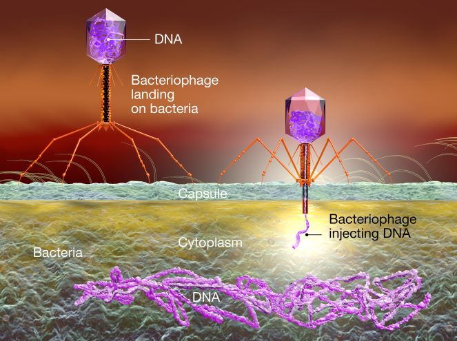
Bacteriophages, also called phages, are the most-abundant organisms in nature. They are viruses that infect bacterial cells, ultimately leading to lysis of the cells and the release of new phage particles.
Figure 1. An artist’s representation of T4 phage infecting a bacterium Image:Anna-Lynn Wegener/Shutterstock.com
Two different activities have been devised using P22 phage (figure 1 and background information): one where the phages are subjected to different temperatures, and another where the phages are subjected to a solution of HCl at different pH values. Both activities are intended as aids to teach protein structure, but they employ different biological principles. They can be used interchangeably if doing both is not possible, or they can both be implemented to teach different principles of protein biology or microbiology.
A suitable age range is about 16 to 18 years old.
Preparing the cultures
The phage stock solution can be prepared well ahead of time: see the phage stock preparation in the supporting material.
Since phages are a kind of virus, they can’t replicate alone; they need a bacterial host. In these activities, we use Salmonella enterica subsp. enterica serovar Typhimurium str. LT2, which is a nonpathogenic strain. Salmonella enterica is the binomial genus and species, Typhimurium is the serovar, and LT2 is the strain.
Note on phage/bacteria combinations
The procedure is based on Salmonella enterica bacteria and P22 phage; however, these experiments can also be done with Escherichia coli and T4 phage.
One to four days before doing the experiments, appropriate bacterial cultures need to be set up. This can be done by the students in an earlier lesson or simply prepared by the teacher or technician.
Safety notes
Bacterial liquid cultures should be treated with bleach before being discarded, and all microorganisms should be discarded into appropriate waste containers.
Materials
Salmonella enterica Typhimurium LT2culture
Lysogeny broth (LB; around 50 ml will be enough for both experiments)
Miller’s LB agar plate
Inoculating loop or sterile cotton swab
Sterile conical tubes or similar
Bunsen/ethanol burner
Sterile conical tubes or similar
Pipettes and pipette tips
Procedure
Salmonella plate
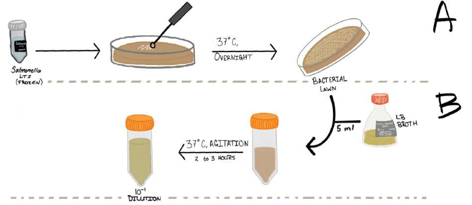
Before the class (1–4 days), take a sample of frozen Salmonella preparation, and use an inoculating loop to streak it over the Miller’s LB agar plate.
Incubate the plate at 37 °C overnight or at room temperature for 24–48 h (figure 2A). The same plate can be used for both activities.
Salmonella liquid culture
Either 3 h before the class or the night before, pipette 5 ml LB broth into a sterile conical tube, and then use the pipette tip to inoculate one colony from the Salmonella culture plate into it.
Incubate the tube with agitation at 37 °C for 3 h or at room temperature without agitation overnight (figure 2B).
From the liquid culture, create a 1/10 dilution using 9 ml LB broth and 1 ml of the liquid culture (figure 2B).
Figure 2. Step-by-step instructions on how to create the bacterial culture plate and the liquid cultures Image courtesy of the author
Activity 1: Heat treatment of bacteriophages
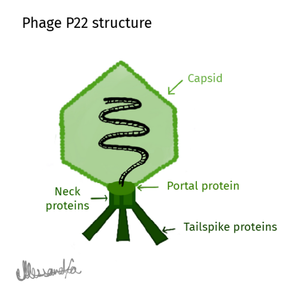
Virtually all known bacteriophages (phages) are composed of a protein capsid and tail, including phage P22. This activity is designed to provide a visual aid to explain this structure, employing the principle of heat denaturation as a basis.
Figure 3. Drawing of the main proteins that make up Lederbergvirus P22 Image courtesy of the author
This activity takes two lessons to complete: one to set up the phage cultures, and a second to evaluate the results after incubation.
Safety notes
Bacterial liquid cultures should be treated with bleach before being discarded, and all microorganisms should be discarded into appropriate waste containers.
10 mM MgSO4 solution (10 ml is enough for both experiments)
TSA culture plates (1-3 per experimental condition)
Liquid agar (3 ml per TSA plate)
Sterile conical tubes or similar
Bunsen/ethanol burner
1.5 ml Eppendorf tubes
Pipettes and pipette tips
Procedure
Hand out the bacteriophage infosheet provided in the supporting material. Explain the structure and main features of phage biology to students before proceeding with the experiments. The students can also view the videos and articles in the resources section or do their own research.
Phage dilution and treatment
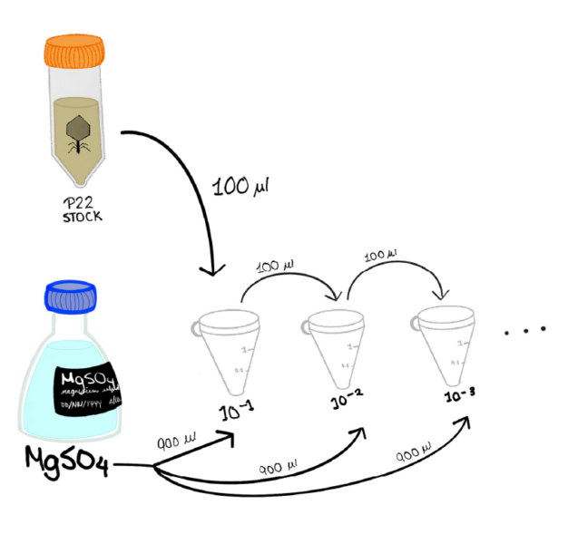
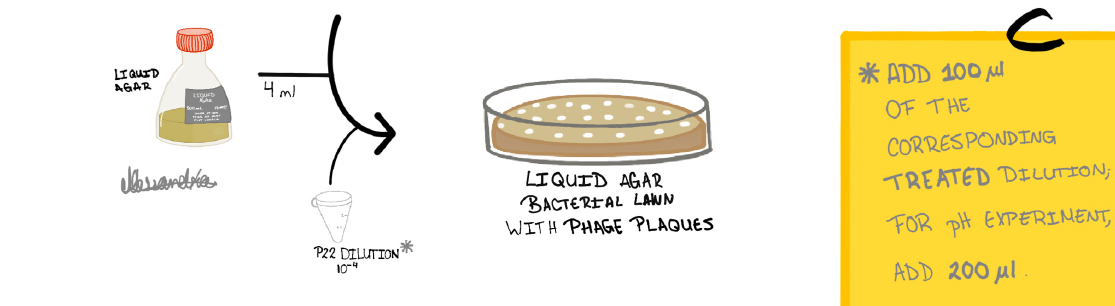
Next, create serial bacteriophage dilutions by taking the 100 µl of phage P22 stock and combining it, in a 1.5 ml Eppendorf tube, with 900 µl of MgSO4 solution. From this first dilution, take 100 µl and combine it in another tube with 900 µl of MgSO4 solution. Repeat twice more to achieve a 10−4 (1/10 000) dilution (figure 4).
Repeat this process to obtain four individual tubes of 10−4 dilutions (figure 4). Dilutions of 10−3 or 10−4 should be suitable, but this may need to be optimized. Ideally, teachers should try the experiment with their phage preparation beforehand, to check the appropriate dilution to give countable plaques.
Figure 4. Step-by-step instructions on creating serial bacteriophage dilutions Image courtesy of the author
Take three of the bacteriophage dilutions and treat them at temperatures higher than 50 °C (recommended: 50 °C, 75 °C, 100 °C) for 5–10 min; the untreated bacteriophage dilution serves as a control. Make sure you label the tubes with the temperature before you start heating.
Phage seeding
Use a permanent marker to label four TSA plates with the three conditions and control. Label the bottom of the plate and not the lid; lids can get mixed up.
Add 3 ml liquid agar to a 15 ml conical tube.
To this tube, add 100 µl of the 1/10 bacterial culture and 100 µl of the untreated 10−4 bacteriophage culture.
Shake by inversion, and pour the mixture over a TSA plate, ensuring it is distributed evenly (figure 5).
Figure 5. Phage seeding Image courtesy of the author
Repeat step 6 for the treated phage dilutions; use one plate for each different condition (temperature). Use several plates for each condition if you want to do statistics, like calculating a mean and standard deviation (SD).
Once all the plates have the corresponding mixture, dispose of the bacterial liquid culture and the bacterial dilution by adding a bleach and mixing; dispose into an appropriate waste container.
Incubate the TSA plates at 37 °C overnight or at room temperature for two days.
Evaluation
After incubation, observe the plates. Is there anything about the plates that becomes more noticeable as the temperature increases?
Ask students to describe what they see. What do they think the clear patches (called plaques) are? Hint: T22 phages are viruses than infect bacteria and destroy them as part of their life cycle.
Having established that the plaques represent phages that infected bacteria and stopped them from multiplying at that point, what does the number of plaques say about the number of infective phages (plaque-forming units) in the sample?
Can students see a difference visually under different temperature conditions?
Optional: the plaques can be counted to create a graph as a visual representation, using the formula:
Results and discussion
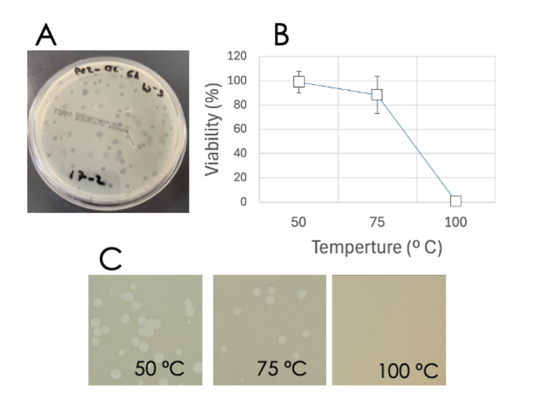
An example of expected results can be seen in figure 6. The control is not shown in this figure, but the incubation temperature can be used and plotted.
Figure 6. Expected results for temperature treatment. A) Image of the plaques obtained in TSA plaques. Effect of temperature on phage viability: B) graph showing the percentage of plaques referenced to an untreated control expressed in mean +/− SD; C) representative images of the plates, showing a decrease in the number of plaques. Image courtesy of the author
Questions to aid the discussion:
Knowing what you now know about bacteriophages, what do you believe this decrease in plaques with temperature is due to?
Can you predict what would happen if we were to use higher temperatures?
Treating proteins at high temperature leads to a phenomenon known as protein denaturation, wherein a protein chain unfolds and the protein loses its 3D shape due to a loss of bonds within the protein. Bacteriophages, being made of proteins, are dependent on the environmental temperature to take on the correct form and be able to infect host cells. In the results, the student will observe how the number of plaques will decrease when the temperature rises, suggesting it is caused by protein denaturation.
Statistics can also be used if several plaques have been seeded for every treatment. The average and SD can be calculated and discussed with the students.
Activity 2: Acidic treatment of bacteriophages
In the next experiment, students can use phages to visualize the effects of suboptimal pH on protein stability.
The activity takes two lessons to complete: one to set up the phage cultures, and a second to evaluate the results after incubation. However, only the treatment of phages is different for the two activities, so the two experiments can be done simultaneously or separately.
Safety notes
Bacterial liquid cultures should be treated with bleach before being discarded, and all microorganisms should be discarded into appropriate waste containers.
Materials
HCl (37%) or HCl solutions at the desired pH (here pH 2 and 4 have been used).
pH strips
100 µl Lederbergvirus/phage P22 stock
MgSO4 10 mM solution (10 ml is enough for both experiments)
TSA culture plates (one per experimental condition)
Liquid agar (3 ml per TSA plate)
Sterile conical tubes or similar
Bunsen/ethanol burner
1.5 ml Eppendorf tubes
Pipettes and pipette tips or measuring glasses
Procedure
Carry out the phage dilution as in steps 1–3 of Activity 1, but only three samples (not four) need to be prepared. Use one for the control and two for testing two different pH (recommended pH 2 and 4).
Instead of heating the samples, take a clean tube and place 100 µl of the desired phage dilution. Add 100 µl of the desired pH HCl solution, leaving it to act for 5 min. Repeat for the two pH conditions. Label the tubes before adding the acid.
Before adding the phages to the agar, use a piece of indicator paper to measure and record the pH of each sample.
Continue with phage seeding and incubation as per steps 5–9 in Activity 1, but use 200 µl of the phage–HCl mix (step 2).
After incubation (37 °C overnight or at room temperature for two days), evaluate the plates and discuss the results as before.
Results and discussion
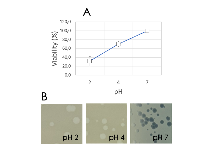
Expected results are shown in figure 7.
Figure 7. Expected results for the effect of pH on phage viability. A) Graph showing the percentage of plaques referenced to an untreated control expressed in mean +/− SD. B) Representative images of the plates obtained. Image courtesy of the author
A decrease in the number of phage plaques can be observed as the pH value decreases, both in the images and in the graph. Proteins exist within a specific pH range: a pH level below or above this pH range will cause the amino acid residues to accept or release protons, which will change their charge and hydrophilicity. This prevents correct folding, which leads to a loss of phage viability. Thus, bacteriophages, being made of proteins, are dependent on the environmental pH to take on the correct structure and be able to enter host cells to replicate themselves.
Questions to aid the discussion:
Is there anything about the plates that becomes more noticeable as the pH becomes more acidic?
Knowing what you now know about bacteriophages, what do you believe this decrease in plaques is due to?
Can you predict what would happen if we were to add an alkali, that is, make the pH more basic?
Conclusion
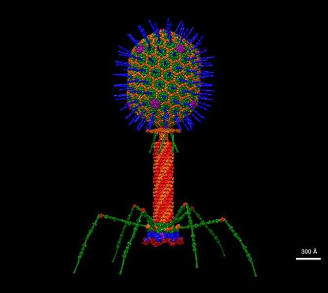
Phages are fascinating organisms and quite engaging, with their weird spaceship structures and the fact that they are viruses that attack bacteria.
Figure 8. Structural model of the T4 phage based on protein structure data Image: Victor Padilla-Sanchez/Wikipedia, CC BY-SA 4.0
The fact that loss of viability leads to the clear and countable visual readout of plaques makes them a good system for demonstrating protein denaturation. Students may also be interested to know that the same plaque-counting approach is used by phage researchers when evaluating different phage mutations or application. In addition to all the discussion possibilities mentioned above, a debate or discussion can be held on whether phages can be used as a treatment against antibiotic-resistant bacteria. This is an obvious question on learning that phages kill bacteria, and if students ask this question, they may be interested to know that phages are indeed being investigated as medical treatments for antibiotic-resistant infections.[1,2] The potential and challenges of this approach are something students could research independently and present to the class.
Understand how phage plaques are formed: Abedon ST (2021) Detection of bacteriophages: phage plaques. In Harper DR et al. (eds) Bacteriophages pp 507–538. Springer. ISBN: 978-3-319-41985-5
Find similar experiments that can be used with students: Richter M, Fraser D (1959) The use of bacteriophages in high school biology. The American Biology Teacher21: 282–287. doi: 10.2307/4439165
Alessandra Mencos Toriello obtained a degree in biochemistry (2024) from the University of Navarra. Currently, she works in protein production using precision fermentation in Portugal and will be pursuing a master’s degree in biomedicine in the future.
Teresa Clemente Galdiano graduated with a degree in biology and business from the University of Navarra in 2023. Currently, she is studying for a master’s degree in infection biology at Uppsala University, Sweden.
Iñigo Izal Azcárate obtained his degree in biochemistry in 1997 and PhD in 2002. For 10 years, he was a researcher in the Orthopaedics Laboratory of the University of Navarra. Currently, he is part of the teaching support staff at the Biochemistry and Genetics Department of the same university.
Camelids are famously robust and useful animals. Surprisingly, their unusual antibodies are just as sturdy and are now revolutionizing medical science.
Biology, Health, Science and society, News from the EIROs
A walk on the wild side: invite some ants to take a walk on your petri dish and discover how bacteria from their feet could help us reduce pesticide use.