
Faster, cheaper, CRISPR: the new gene technology revolution
A controversial new technology is making gene editing far cheaper and easier – too easy,…
Understand, Biology





Read about discovery of light-activated molecular switches that have provided the much-needed tools for taking neuroscience and biology research to the next level.
This year’s Nobel Prize in Physiology or Medicine started with the discovery of a special light-sensing protein and engineered it into a tool that has moved our understanding of our own brain and biology by leaps and bounds. On 10 December 2026, Karl Deisseroth, Peter Hegemann and Georg Nagel will be presented with the most prestigious award in science for their development of optogenetics.[1] As they are preparing for their flight to Stockholm, we get to learn more about what it means to use light as a switch for cells.

Light is all around us, and sometimes we forget how cool it is that we can detect it. The retina in our eyes is filled with cells that are activated in response to light, aptly called photoreceptor cells. Photoreceptor cells can do this because their cell membranes have special proteins called opsins. When hit by light, an opsin protein changes its shape and triggers a chain reaction in a cell.[2] The chain reaction causes the cell to generate an electrical signal, which is sent to the brain, et voilà – you can read this article.
Being fascinated by their ability to change shape in response to light, scientists have been studying opsins since the 1970s. There are many different opsins found in nature.[3] In addition to our eyes, many unicellular organisms, such as bacteria and algae, have opsins, which they use to guide their movements in response to light or to produce energy. The details of cellular changes depend on the opsin type and the organism, but bacterial opsins share a basic mechanism with those in our eyes. Opsins change their shape in response to incident light, opening channels that let many charged particles enter the cell. Changes in the cell’s electric potential signal it to move in a specific direction. The movement of charged particles can also be linked to the energy-producing reactions in the cell.[4,5] Nevertheless, the opsins found in these unicellular organisms differ from the opsins in our eye in a very fundamental way, which proved to be pivotal in the discovery of optogenetics.
In the 1990s, Peter Hegemann’s team showed that opsins drive light-directed movement in Chlamydomonas, a unicellular alga (figure 1).[6] This specific opsin, called channelrhodopsin-2, is different from the ones found in our eyes.
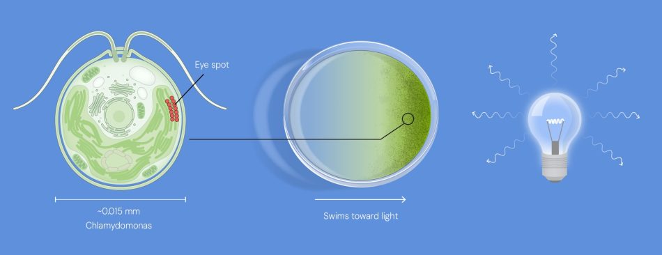
Channelrhodopsin-2 is not only a light detector, but also a gate that opens in response to light. When light falls on the protein, it opens its channel and lets positively charged particles (protons or cations) into the cell. The change in concentration of electrically charged particles causes the cell to activate.[4,5] This reaction is very fast and precise, making it highly relevant for quick changes in response to light.
Following this discovery, Hegemann established a collaboration with Georg Nagel, who had experience in studying ion channels such as channelrhodopsins. Utilising the power of genetic engineering, Georg Nagel and his colleagues expressed channelrhodopsin-2 in frog egg cells and then in human kidney cells in the early 2000s.[7] These initial studies established that genetically engineered channelrhodopsins can indeed be expressed in other cell types, allowing scientists to develop a system that can change the cellular electric potential within milliseconds using light. Given that electrical signals are the basis of our brain function, these findings inspired Karl Deisseroth to develop the same system to work in neurons.
Simply because we know very little about our brain. Neurons are the cells that transmit signals and process all the information inside our brains. The interaction between neurons is how we learn, feel, move and experience the world around us. For years, neuroscientists have tried to figure out the details of these interactions, but their understanding has remained vague because they did not have the tools to capture them.[8] Peeking into an active brain to look at a specific neuronal circuit is tricky because a lot is going on in the brain at the same time. To establish proof that specific neurons in a region cause specific behaviour, we need to observe the effect of rapidly activating and silencing them.
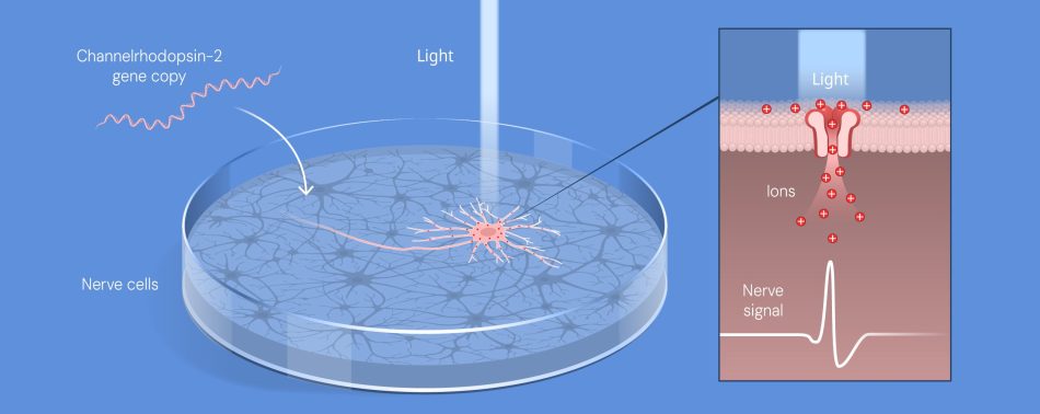
The challenge of activating neurons using an opsin from a completely different organism was a big one for Karl Diesseroth and his team.[9] After much trial and error, tweaking methods, and optimising the molecule itself, Karl and his colleagues were able to express channelrhodopsin-2 in neurons grown on a petri dish.[10] Their study showed that light-based modulation of channelrhodopsin-2 can activate neurons and established the field of ‘optogenetics’, which uses genetically modified proteins that can be switched on or off using light to control cells (figure 2).
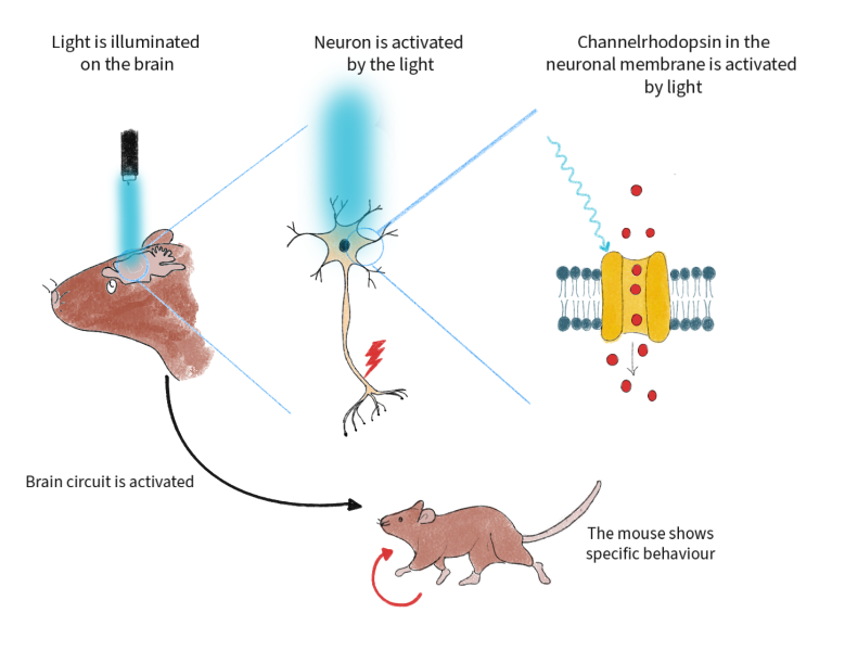
Eventually, many other neuroscientists around the world placed channelrhodopsin-2 in the neurons of different model organisms, including chickens,[11] worms[12] and flies,[13] to show that they could control specific behaviours in animals by switching neurons on and off with light (figure 3). Scientists have also discovered and designed different variants of opsins to target specific types of neurons, brain regions and functions, enabling them to study these areas more effectively.[14–16] Within the first three years of inserting channelrhodopsin-2 into neurons, scientists began to unravel the neuronal mechanisms behind complex functions such as learning, sensations, sight, breathing and movement. These findings fed directly into the study of psychological disorders, including depression, schizophrenia and anxiety, as well as neurodegenerative disorders such as Parkinson’s disease.[17]
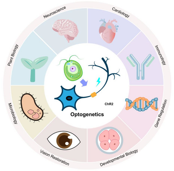
While neuroscience remains at the forefront of optogenetics, it has also benefited the study of heart muscle cells, advancing possible interventions for cardiac arrest and heart failure.[18] The ability to exert highly precise control at molecular-scale and at milliseconds rates allows optogenetics to be a multifaceted tool that is also used in immunology, metabolism, plant biology, development and cancer research.[19,20]
One of the biggest achievements in clinical application of optogenetics emerged in 2021. Optogenetics was used to partially restore vision for a patient with retinitis pigmentosa, an eye disease in which loss of light-sensing receptors can cause vision loss.[21] Within two decades, optogenetics went from briefly activating some neurons in a petri plate to restoring vision. This exemplifies how curiosity-driven interdisciplinary research forms the basis for advanced applications that can improve lives.[17] Optogenetics is now moving towards the development of more precision, integration with newer genetic advancements such as the gene editing technique CRISPR and diversification of applications in plant biology, cancer research and larger-scale clinical trials. The field also needs to overcome many challenges that are inherent in using light and genetics. In animal models, optogenetics can only be used for organs that can be penetrated by light, unless invasive surgery is involved. Moreover, the introduction of foreign proteins and genes can trigger immune responses and off target effects.[17] Yet we can rest assured that through the sheer desire to solve problems and answer fundamental questions, the field of optogenetics will keep moving forward.
[1] Press release of the Nobel Prize in Physiology or Medicine 2026: https://www.nobelprize.org/prizes/medicine/2026/press-release/
[2] Shichida Y, Matsuyama T (2009) Evolution of opsins and phototransduction. Philosophical Transactions of the Royal Society B: Biological Sciences 364: 2881–2895. doi: 10.1098/rstb.2009.0051
[3] Friedman JM (2021) How the discovery of microbial opsins led to the development of optogenetics. Cell 184: 5266–5270. doi: 10.1016/j.cell.2021.08.022
[4] Harz H, Hegemann P (1991) Rhodopsin-regulated calcium currents in Chlamydomonas. Nature 351: 489–491. doi: 10.1038/351489a0
[5] Spudich JL, Lanyi JK (1996) Shuttling between two protein conformations: the common mechanism for sensory transduction and ion transport. Curr. Opin. Cell Biol. 8: 452–457. doi: 10.1016/S0955-0674(96)80020-2
[6] Kateriya S et al. (2004) “Vision” in Single-Celled Algae. News Physiol Sci 19: 133–137. doi: 10.1152/nips.01517.2004
[7] Hegemann P, Nagel G (2013) From channelrhodopsins to optogenetics. EMBO Mol. Med. 5: 173. doi: 10.1002/emmm.201202387
[8] The science behind optogentics: https://www.nobelprize.org/uploads/2026/10/advanced-medicineprize2026.pdf
[9] Boyden ES (2011) A history of optogenetics: the development of tools for controlling brain circuits with light. F1000 Biol. Rep. 3: 11 doi: 10.3410/B3-11
[10] Boyden ES et al. (2005) Millisecond-timescale, genetically targeted optical control of neural activity. Nature Neuroscience 8: 1263–1268. doi: 10.1038/nn1525
[11] Inaba M, Shikaya Y, Takahashi Y (2026) Optogenetics as a useful tool to control excitable and non-excitable tissues during chicken embryogenesis. Dev. Biol. 529: 81–87. doi: 10.1016/j.ydbio.2025.10.004
[12] Nagel G et al. (2005) Light activation of Channelrhodopsin-2 in excitable cells of caenorhabditis elegans triggers rapid behavioral responses. Current Biology 15: 2279–2284. doi: 10.1016/j.cub.2005.11.032
[13] Schroll C et al. (2006) Light-Induced Activation of Distinct Modulatory Neurons Triggers Appetitive or Aversive Learning in Drosophila Larvae. Current Biology 16: 1741–1747. doi: 10.1016/j.cub.2006.07.023
[14] Dawydow A et al. (2014) Channelrhodopsin-2-XXL, a powerful optogenetic tool for low-light applications. Proc. Natl. Acad. Sci. U. S. A. 111: 13972–13977. doi: 10.1073/pnas.1408269111
[15] Klapoetke NC et al. (2014) Independent optical excitation of distinct neural populations. Nature Methods 11: 338–346. doi: 10.1038/nmeth.2836
[16] Prakash R et al. (2012) Two-photon optogenetic toolbox for fast inhibition, excitation and bistable modulation. Nature Methods 9: 1171–1179. doi: 10.1038/nmeth.2215
[17] Duan X, Zhu M, Gao S (2025) Two Decades of Optogenetic Tools: A Retrospective and a Look Ahead. Advanced Genetics 6: e00021
[18] Boyle PM, Karathanos, TV, Trayanova NA Cardiac (2018) Optogenetics: 2018. JACC Clin. Electrophysiol. 4: 155–167. doi: 10.1002/ggn2.202500021
[19] Emiliani V et al. (2022) Optogenetics for light control of biological systems. Nature Reviews Methods Primers 2. doi: 10.1038/s43586-022-00136-4
[20] Krueger D et al. (2019) Principles and applications of optogenetics in developmental biology. Development (Cambridge) 146. doi: 10.1242/dev.175067
[21] Sahel JA et al. (2021) Partial recovery of visual function in a blind patient after optogenetic therapy. Nat. Med. 27: 1223–1229. doi: 10.1038/s41591-021-01351-4
A controversial new technology is making gene editing far cheaper and easier – too easy,…
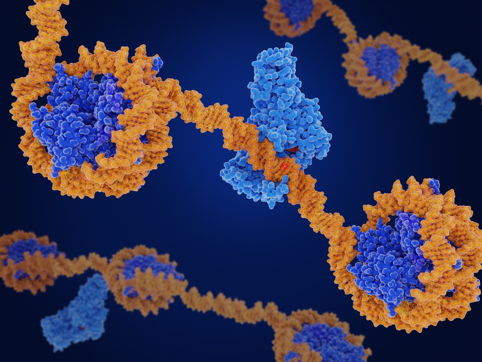
Our DNA is the blueprint of life, but epigenetics interprets how it is read. Learn how epigenetics controls our genes, enables life and shapes who we are.
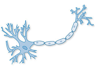
Simulate a neuron in the…

